Inside the Scan
Focused cardiac imaging for complete reassurance
A dedicated fetal echocardiogram examines the heart's structure and function in detail, using imaging tuned specifically for fetal cardiac assessment.
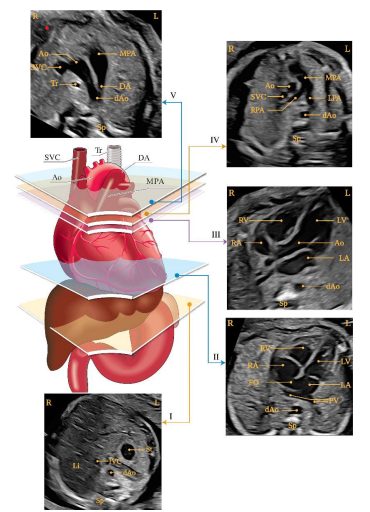
Fetal Echocardiography is a specialised scan that focuses only on your baby’s heart - evaluating its structure, chambers, valves and blood flow in fine detail, performed with FMF and ISUOG-aligned expertise.
A dedicated fetal echocardiogram examines the heart's structure and function in detail, using imaging tuned specifically for fetal cardiac assessment.
Fetal echocardiography is a focused, high-resolution ultrasound examination of the fetal heart. It is typically performed between 22 and 24 weeks, and evaluates the four heart chambers, major vessels, valves and blood flow patterns to detect congenital heart conditions early - allowing families and care teams to plan ahead with clarity.
A fetal heart scan is recommended when there is a higher chance of a heart condition, or as a specialist confirmation after a routine scan finding.
Recommended when there is a family history of congenital heart disease.
Advised for pregnancies with pre-existing or gestational diabetes.
Performed as a specialist follow-up after a suspected finding on a routine or NT scan.
Recommended when NT measurement or genetic screening indicates higher risk.
It is typically performed between 22 and 24 weeks of pregnancy, though it can sometimes be done earlier in specialist settings.
Common reasons include a family history of heart conditions, maternal diabetes, an increased NT measurement, or a suspected finding on a routine anomaly scan.
Yes - it uses standard diagnostic ultrasound, the same safe technology used in routine pregnancy scans, with no known risk to mother or baby.
You will receive a clear explanation of the finding, its implications, and a coordinated care plan - which may include closer monitoring, paediatric cardiology input, and delivery planning at an appropriate facility.
Call, WhatsApp, or request an appointment online - our care team will confirm your slot with Dr. Priyanka.